Diagnosis of Chronic Cholecystitis
The diagnosis of chronic cholecystitis can be suspected if the patient complains of having dull or acute pain in the right hypochondrium as a result of eating fatty or fried food, drinking soft drinks, and other foods and beverages that stimulate the production of bile. However, the diagnosis can only be verified using specific diagnostic tools including:
Visualization methods
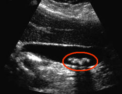
- Abdominal ultrasound – this is the first method that is used to diagnose chronic cholecystitis. Ultrasound can clearly show the inflammatory process within the gallbladder (its walls become thicker) and the gallstones (which are usually the cause or the result of the chronic cholecystitis). Moreover, this diagnostic tool is quite cheap, so any patient can afford to go through it.
- Abdominal CT scan – just like the ultrasound, CT scan can detect gallstones within the gallbladder and assess its condition. In comparison to ultrasound, computed tomography is more sensitive in detecting pericholecystis inflammatory response and localizing pericholecystic gas, pericholecystic abscesses, and gallstones that are not located within the gallbladder.

- Hepatobiliary scintigraphy using technetium-99m is also an accurate and sensitive method of diagnosing chronic cholecystitis. This method uses nuclear imaging to detect gallstones and assess the function of the gallbladder. If the ejection fraction of the gallbladder is low (a small amount of bile escapes the gallbladder), this can indicate a chronic cholecystitis. In order to perform this method, a radioactive tracer (usually Tc-iminodiacetic acid chelate complex) has to be injected into a vein. It is then excreted by the biliary system and stored within the gallbladder. If all is well the gallbladder can be visualized after 1 hour after the injection. However, if there is some form of obstruction, the gallbladder might not show up on the scan even within 4 hours. Cholescintigraphy has a higher sensitivity than ultrasonography and has the ability to differentiate between Biliary atresia (a congenital or acquired condition during which the common bile duct is missing) and Neonatal Hepatitis (an inflammation of liver’s tissues that occurs in early infancy).
Additional methods
- Liver functions tests – these tests can identify the levels of direct and indirect bilirubin, alkaline phosphatase and liver transaminases (AST and ALT). Usually, chronic cholecystitis doesn’t cause a rise in these parameters. However, if something obstructs the common bile duct, it can lead to increased levels of bilirubin, alkaline phosphatase, and transaminases.
- Complete blood count – a rise in white blood cells can indicate the chronic disease has “flared-up.” Usually this happens as a result of a mistake in diet.

- Levels of pancreatic enzymes – when diagnosing chronic cholecystitis it is important to exclude pancreatitis as the cause of the pain. Raised levels of lipase and amylase can indicate that the pain might be caused by a flare-up of chronic pancreatitis.