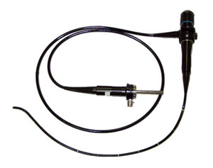
Endoscopy

This method is based on the examination of the internal organs from the inside using a special optical device called an endoscope. This instrument is usually inserted into the patient through his natural ways, for example, into the stomach through the mouth, into the straight intestine (rectum) – through the anus. However, sometimes punctures in the abdominal wall can be used for this purpose.
The diameters of endoscopes may vary from 0.8 cm to 1.5 cm and the length – from 30 cm to 1.5 m. Fiber-optic video systems make endoscope flexible and provide the ability to have the image on the screen. Many endoscopes are equipped with a device that can be used to take tissue samples and an electrical probe to destroy pathological tissues.
The endoscopemakes it possible to get a clear image of the mucous membrane of the digestive tract, to see ulcers, the areas of irritation, inflammation and pathological growth of tissuesas well as to take samples for the further analysis. The endoscope can also be used for treatment purposes.
In each particular case, endoscopy is performed with the help of a special endoscope slightly different in structure in accordance with the anatomical-physiological peculiarities of the organ under observation. The procedures are named for the organ being examined:
- Gastroscopy - endoscopic study of the stomach
- Esophagoscopy- endoscopic examination of the esophagus
- Duodenoscopy - endoscopic study of the duodenum
- Colonoscopy- endoscopic examination of the large intestine (colon)
- Proctoscopy- endoscopic study of the rectum and sigmoid colon
